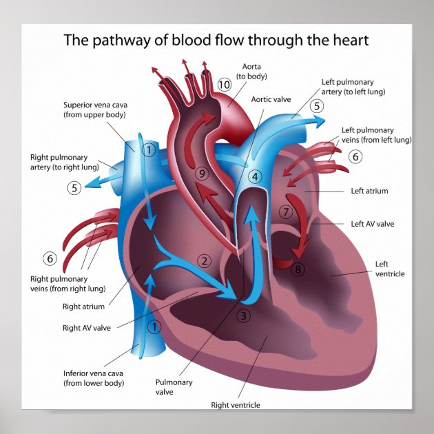
Ĭardiovascular diseases (CVD) are the most common cause of death globally as of 2008, accounting for 30% of deaths. Exercise temporarily increases the rate, but lowers resting heart rate in the long term, and is good for heart health. The heart beats at a resting rate close to 72 beats per minute. Oxygenated blood then returns to the left atrium, passes through the left ventricle and is pumped out through the aorta into systemic circulation, traveling through arteries, arterioles, and capillaries-where nutrients and other substances are exchanged between blood vessels and cells, losing oxygen and gaining carbon dioxide-before being returned to the heart through venules and veins.

From here it is pumped into pulmonary circulation to the lungs, where it receives oxygen and gives off carbon dioxide. In humans, deoxygenated blood enters the heart through the right atrium from the superior and inferior venae cavae and passes it to the right ventricle. These generate a current that causes the heart to contract, traveling through the atrioventricular node and along the conduction system of the heart. The heart pumps blood with a rhythm determined by a group of pacemaker cells in the sinoatrial node. The wall of the heart is made up of three layers: epicardium, myocardium, and endocardium. The heart is enclosed in a protective sac, the pericardium, which also contains a small amount of fluid. In a healthy heart blood flows one way through the heart due to heart valves, which prevent backflow. Fish, in contrast, have two chambers, an atrium and a ventricle, while most reptiles have three chambers. Commonly the right atrium and ventricle are referred together as the right heart and their left counterparts as the left heart. In humans, other mammals, and birds, the heart is divided into four chambers: upper left and right atria and lower left and right ventricles. In humans, the heart is approximately the size of a closed fist and is located between the lungs, in the middle compartment of the chest. The pumped blood carries oxygen and nutrients to the body, while carrying metabolic waste such as carbon dioxide to the lungs. This organ pumps blood through the blood vessels of the circulatory system. Atherosclerosis (a buildup of plaque in the inner lining of an artery causing it to narrow or become blocked) is the most common cause of heart disease.The heart is a muscular organ in most animals. This can lead to a heart attack and possibly death. Since coronary arteries deliver blood to the heart muscle, any coronary artery disorder or disease can have serious implications by reducing the flow of oxygen and nutrients to the heart muscle. Smaller branches of the coronary arteries include: obtuse marginal (OM), septal perforator (SP), and diagonals. Together with the left anterior descending artery, the right coronary artery helps supply blood to the middle or septum of the heart. The right coronary artery divides into smaller branches, including the right posterior descending artery and the acute marginal artery. The right coronary artery supplies blood to the right ventricle, the right atrium, and the SA (sinoatrial) and AV (atrioventricular) nodes, which regulate the heart rhythm. This artery supplies blood to the outer side and back of the heart. The circumflex artery branches off the left coronary artery and encircles the heart muscle. The left anterior descending artery branches off the left coronary artery and supplies blood to the front of the left side of the heart. The left main coronary divides into branches: The left main coronary artery supplies blood to the left side of the heart muscle (the left ventricle and left atrium). The 2 main coronary arteries are the left main and right coronary arteries. What are the different coronary arteries? Small branches dive into the heart muscle to bring it blood. The coronary arteries wrap around the outside of the heart. Also, oxygen-depleted blood must be carried away. Like all other tissues in the body, the heart muscle needs oxygen-rich blood to function.

Coronary arteries supply blood to the heart muscle.


 0 kommentar(er)
